Atrial tachycardia
http://emedicine.medscape.com/article/151456-overview
Atrial tachycardia is a rhythm disturbance that arises in the atria. Atrial tachycardia can be observed in persons with normal hearts and in those with structurally abnormal hearts, including those with congenital heart disease and particularly after surgery for repair or correction of congenital or valvular heart disease.
Atrial tachycardia is defined as a supraventricular tachycardia (SVT) that does not require the atrioventricular (AV) junction, accessory pathways, or ventricular tissue for initiation and maintenance of the tachycardia. In common with most of the SVTs, the ECG typically shows a narrow QRS complex tachycardia (unless bundle branch block aberration occurs). Heart rates during atrial tachycardia are highly variable, with a range of 100-250 beats per minute (bpm). The atrial rhythm is usually regular. The conducted ventricular rhythm is also usually regular but may become irregular, often at higher atrial rates because of variable conduction through the AV node, thus producing conduction patterns such as 2:1, 4:1, a combination of those, or Wenckebach AV block.
The P wave morphology as observed on the ECG may give clues to the site of origin and mechanism of the atrial tachycardia. In the case of a focal tachycardia, the P wave morphology and axis depend on the location in the atrium from which the tachycardia originates. In the case of macroreentrant circuits, the P wave morphology and axis depend on activation patterns (see Workup).
Multifocal atrial tachycardia (MAT) is an arrhythmia with an irregular atrial rate greater than 100 bpm. Atrial activity is well organized, with at least 3 morphologically distinct P waves, irregular P-P intervals, and an isoelectric baseline between the P waves. Shine et al first proposed this definition in 1968.[1] Multifocal atrial tachycardia has previously been described by names such as chaotic atrial rhythm or tachycardia, chaotic atrial mechanism, and repetitive paroxysmal MAT. Go to Multifocal Atrial Tachycardia for more complete information on this topic.
A number of methods are used to classify atrial tachycardia, including origin as based on endocardial activation mapping data, pathophysiologic mechanisms, and anatomy.
Based on endocardial activation, atrial tachycardia may be divided into 2 groups. The first is focal atrial tachycardia, which arises from a localized area in the atria such as the crista terminalis, pulmonary veins, ostium of the coronary sinus, or intra-atrial septum. The second group is the reentrant atrial tachycardias. These reentrant (usually macroreentrant) atrial tachycardias most commonly occur in persons with structural heart disease, complex heart disease, and particularly after surgery involving incisions or scarring in the atria (see Clinical Presentation).
Atrial tachycardia may be classified according to the following pathophysiologic mechanisms: enhanced automaticity, triggered activity, or reentry (see Pathophysiology). Anatomical classification of atrial tachycardia is based on the location of the arrhythmogenic focus (see Anatomy).
A 12-lead electrocardiogram (ECG) is an important tool to help identify, locate, and differentiate atrial tachycardia. Laboratory studies may be indicated to exclude systemic disorders that may be causing the tachycardia. Electrophysiologic study may be required (see Workup).
The primary treatment during a bout of atrial tachycardia is considered to be rate control using AV nodal blocking agents, such as beta-blockers or calcium channel blockers (see Medication). Cardioversion should be considered for any patient in whom the rhythm is not tolerated well hemodynamically and in whom rate control drugs are ineffective or contraindicated. Radiofrequency catheter ablation for atrial tachycardia has become a highly successful and effective treatment option for symptomatic, medically refractory patients or those who do not desire long-term antiarrhythmic therapy. (see Treatment and Management.)
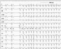 This 12-lead electrocardiogram demonstrates an atrial tachycardia at a rate of approximately 150 beats per minute. Note the negative P waves in leads III and aVF (upright arrows) are different from the sinus beats (downward arrows). The RP interval exceeds the PR interval during the tachycardia. Note also that the tachycardia persists despite the atrioventricular block.
 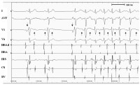 Note that the atrial activities originate from the right atrium and persist despite the atrioventricular block. These features essentially exclude atrioventricular nodal reentry tachycardia and atrioventricular tachycardia via an accessory pathway. Note also that the change in the P wave axis at the onset of tachycardia makes sinus tachycardia unlikely.
 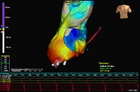 Anterior-posterior projection is shown. An example of activation mapping using contact technique and EnSite system. The atrial anatomy is partially reconstructed. Early activation points are marked with white/red color. The activation waveform spreads from the inferior/lateral aspect of the atrium thought the entire chamber. White points indicate successful ablation sites that terminated the tachycardia. TV – Tricuspid valveCS – Shadow of the catheter inserted in the coronary sinus
 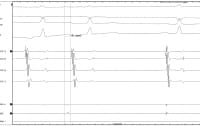 Intracardiac tracings showing atrial tachycardia breaking with application of radiofrequency energy. The local electrograms in the successful site preceded the surface P wave by 51 ms, consistent with successful site. Note that postablation electrograms on the ablation catheter is inscribed well past the onset of sinus rhythm P wave. The first 3 tracings show surface electrocardiograms as labeled.CS – Respective pair of electrodes of the coronary sinus catheterCS 7,8 – Located at the os of the coronary sinusCS 1,2 – Distal pair of electrodes Abl – Ablation catheter (D-distal pair of electrodes)
 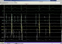 An example of rapid atrial tachycardia mimicking atrial flutter. Single radiofrequency application terminates the tachycardia. The first 3 tracings show surface electrocardiograms, as labeled. HRA – High right atrial catheterRVA – Catheter located in right ventricular apexHBED and HBEP – Respectively, distal and proximal pair of electrodes in the catheter located at His bundleAblD and AblP – Respectively, distal and proximal pair of electrodes of the mapping catheterMAP – Unipolar electrograms from the tip of the mapping catheter
  خوشبختى لذت مشتركى است كه حاصل يارى بى چشمداشت به ديگران است .